.jpg)
What to Expect During a Dental X-Ray Near Me
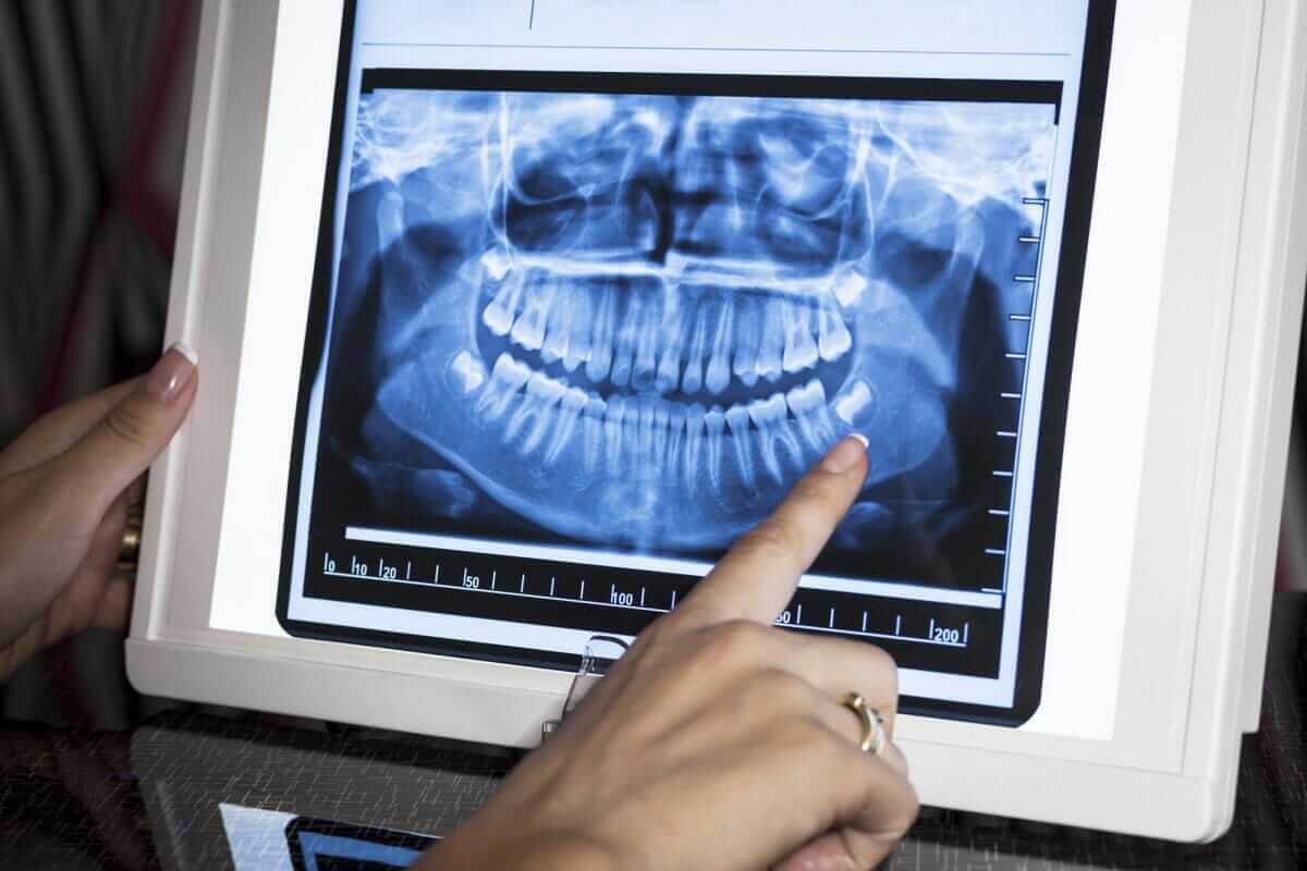
What to Expect From a Dental X-Ray in Urbana
If you find yourself searching for a dental X-ray near me, finding a warm and welcoming office makes the process easy. Start by scheduling a dentist appointment for an examination. Your dentist can check whether you need updated imaging and choose the exact views required. During routine dentist teeth cleaning visits, bitewing X-rays help spot early decay between teeth, while other views reveal tooth roots, jawbone health, or developing teeth.
During your visit for trusted dental care near me, a team member places a small digital sensor in your mouth and captures clear images in seconds. The imaging itself does not hurt, though the sensor may feel awkward for a brief moment. At Champaign Dental Group in Urbana, we take time to explain why each image is needed and walk you through what the results show.
I'm Dr. Ellen Zwick, and I'll explain what happens next, including safety protocols, recommended schedules, and the types of images your dentist may suggest.
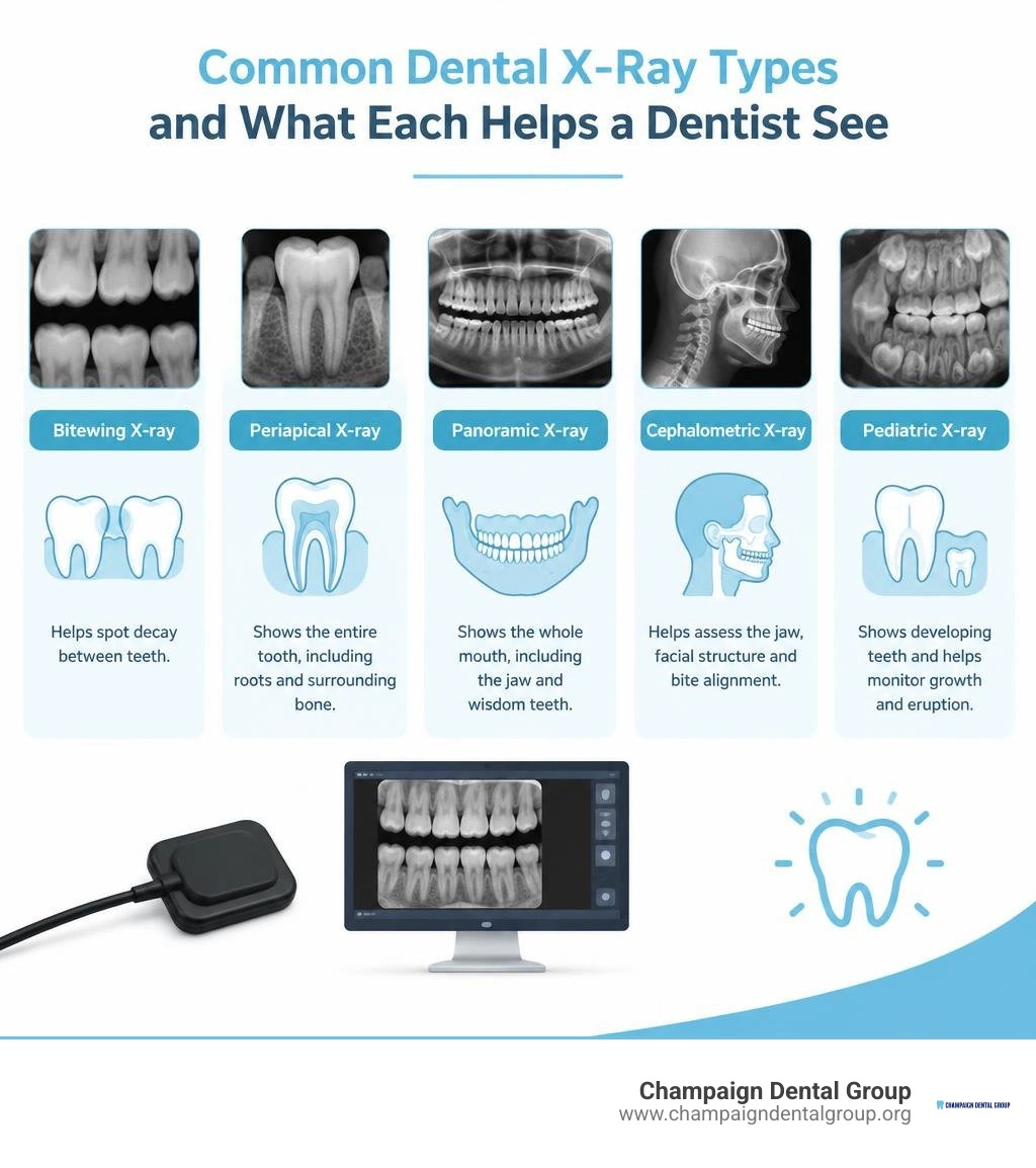
Understanding Diagnostic Imaging: Types of Dental Radiographs
Visual dental checks give us a clear view of your enamel surfaces and gum tissues. However, a significant portion of your oral anatomy stays hidden beneath the gumline and inside the jawbone. Diagnostic dental imaging lets our team see what is happening beneath the surface, identifying concerns before they cause pain.

By taking detailed radiographs, we can evaluate interproximal spaces where adjacent teeth meet, check the alveolar bone that anchors your teeth, and ensure developing structures are healthy.
Intraoral Radiographs: Bitewing, Periapical, and Occlusal Views
Intraoral radiographs are images taken with the sensor placed gently inside your mouth. These are the most common views used in routine preventative dentistry care.
- Bitewing Radiographs: These images focus on the crowns of your upper and lower back teeth in a single view. They are the gold standard for spotting hidden decay between teeth and monitoring early bone level changes caused by gum disease.
- Periapical Views: A periapical image captures the entire tooth from the chewing surface down to the root tip and surrounding jawbone. We rely on these views to evaluate deep infections, abscesses, or changes in the supporting bone.
- Occlusal Imaging: Larger than standard intraoral views, occlusal radiographs highlight the floor or roof of your mouth. They help track full arches, pinpoint extra teeth, locate impacted teeth, or identify jaw abnormalities in children.
Extraoral and 3D Imaging: Panoramic and CBCT Scans
Extraoral images use sensors positioned outside the mouth. These systems capture wide views of the head, jaw, and facial structures, making them especially comfortable for patients with a sensitive gag reflex.
- Panoramic Radiographs: A panoramic scan moves around the outside of your head to provide a continuous, ear-to-ear overview of your entire mouth. It displays all upper and lower teeth, the maxillary sinuses, and the temporomandibular joints (TMJ) in a single image.
- Cephalometric Projections: These scans show the entire side profile of the head and face. Orthodontists and dentists use them to evaluate jaw growth and plan bite corrections.
- Cone Beam Computed Tomography (CBCT): CBCT systems take dental imaging a step further by generating 360-degree, three-dimensional volumetric scans. Advanced systems capture high-resolution anatomical views in as little as 14 seconds. These detailed 3D views are vital for airway evaluation, complex root canal therapy, and precise implant planning. For patients considering implants, reviewing scientific research on dental implants and bone integration highlights why knowing exact bone density in advance is crucial for lasting stability.
Radiation Safety and Modern Digital Radiography
It is natural to wonder about radiation safety whenever an imaging procedure is recommended during a comprehensive dental examination. In modern dental practices, patient safety is guided by the ALARA principle, which stands for "As Low As Reasonably Achievable." We use modern protocols and equipment to ensure you receive the lowest diagnostic exposure possible.
Radiation Exposure Levels and Protective Protocols
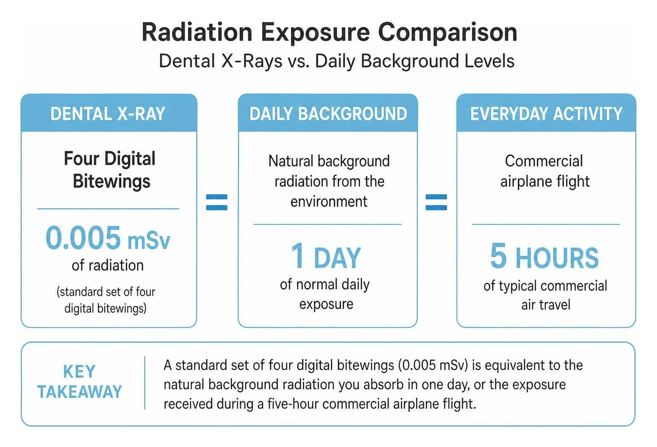
Dental radiographs use remarkably small amounts of radiation. A standard set of four digital bitewings exposes you to roughly 0.005 millisieverts (mSv) of radiation. To put that in perspective, that is equivalent to the natural background radiation you absorb from the environment in a single normal day.
During a typical dental imaging session, your total exposure is comparable to what you would naturally receive on a five-hour commercial airplane flight. We also use protective equipment, such as lead aprons with contoured thyroid collars and focused machine collimators, to shield the rest of your body and keep radiation focused only on the necessary area.
Key Advantages of Digital Imaging Over Traditional Film
The dental field has largely moved away from traditional film packets, and modern digital radiography offers major benefits for patient health and comfort:
- Up to 80% to 90% Radiation Reduction: Digital sensors require far less radiation to produce crisp, high-detail images than old-fashioned chemical film.
- Instant Image Display: Digital images appear on chairside computer monitors within seconds, eliminating long waits for darkroom processing.
- Enhanced Diagnostic Clarity: We can adjust contrast, zoom in on suspicious areas, and show you exactly what we see on screen.
- Eco-Friendly Care: Digital systems eliminate the need for harsh chemical fixers and developers, reducing environmental waste.
- Secure Record Keeping: Digital radiographs integrate directly with secure electronic health records, making them easy to track over time or share when coordinated care is required.
Step-by-Step: What to Expect During Your Dental Imaging Appointment
If you feel nervous about having X-rays taken, knowing the workflow ahead of time can help you feel completely at ease. The entire process is non-invasive, quick, and comfortable.
What Happens During a Routine Dental X-Ray Near Me?
When you arrive for your visit, our team ensures you are seated comfortably in the dental chair:
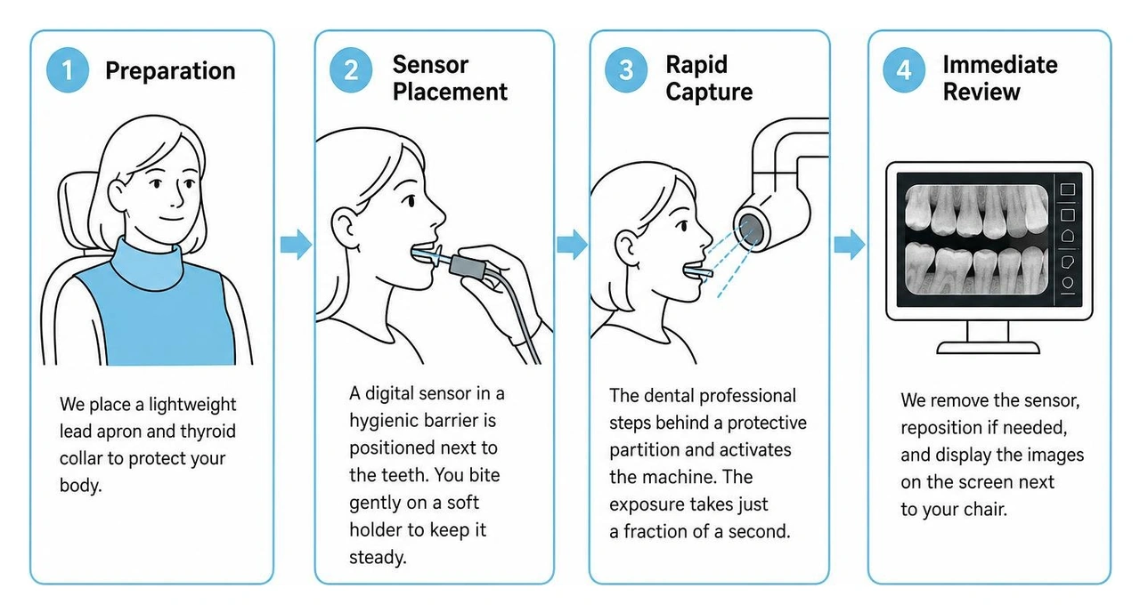
- Preparation: We place a lightweight lead apron and thyroid collar over your chest and neck to protect your body.
- Sensor Placement: A small, smooth digital sensor encased in a hygienic barrier is gently positioned next to the teeth being imaged. You will bite down gently on a soft plastic holder to keep the sensor steady.
- Rapid Capture: The dental professional steps behind a protective partition and activates the machine. The exposure takes just a fraction of a second.
- Immediate Review: We remove the sensor, reposition it if additional angles are needed, and immediately display the completed images on a screen next to your chair.
There is zero pain involved. You can ask questions, look at your teeth on the monitor, and discover why the importance of regular dental check-ups beyond cleaning keeps your mouth healthy year after year.
Advanced 3D Cone Beam CT (CBCT) Imaging Experience
If your treatment calls for a 3D scan, the process is even simpler. You do not need to hold sensors inside your mouth. Instead, you stand or sit upright in an open machine while resting your chin on a comfortable support. The machine's rotating arm circles smoothly around your head for less than a minute. The system captures multi-angle views and reconstructs them into an interactive 3D model of your jaw, roots, and facial bones.
Recommended Frequency for Dental Radiographs
Dentists do not take X-rays on a rigid, one-size-fits-all timeline. Instead, we customize imaging schedules based on your individual risk factors, past dental history, and current oral health status. Consistent check-ups offer several advantages, as outlined in our guide on the 7 benefits of having a regular dental check up.
How Often Should I Schedule a Dental X-Ray Near Me?
- Healthy Adults with Low Cavity Risk: Routine bitewing X-rays are generally recommended once every one to two years, while a comprehensive full-mouth series or panoramic scan is updated every three to five years.
- Adults with High Cavity or Gum Disease Risk: If you have active decay, deep gum pockets, or extensive restorations, your dentist may suggest bitewings every six to twelve months to catch changes early.
- Teenagers: Because teeth and jaws are still growing and wisdom teeth may be forming, teenagers often receive bitewing X-rays every one-and-a-half to three years, alongside periodic panoramic views.
Special Considerations for Children and Pregnant Patients
Children have developing teeth with thinner enamel, which makes them more vulnerable to rapid tooth decay. For kids at low risk of cavities, bitewing images are recommended every one to two years. Pediatric digital sensors use ultra-low radiation settings designed specifically for growing bodies.
If you are pregnant or think you might be, let your dental team know right away. Routine preventive X-rays can easily be postponed until after delivery. However, if a dental infection or emergency arises, diagnostic X-rays can be performed safely during pregnancy with double-layer lead aprons to shield your baby.
How to Choose the Right Provider for a Dental X-Ray Near Me
When searching for trusted dentists in Champaign County, look for a practice that pairs modern technology with warm, individualized attention. High-quality imaging equipment makes your appointments faster and more informative, but gentle, attentive care is what makes you feel comfortable in the chair.
At Champaign Dental Group, we have cared for our neighbors as a trusted Champaign County staple since 1997. We treat every patient like family, welcoming children, parents, and seniors from Urbana, Mechanicsburg, Tremont City, West Liberty, and Springfield. Our team provides comprehensive oral health care, ranging from routine cleanings to in-house oral surgery and sedation dentistry for individuals with dental anxiety.
To help make exceptional care accessible, we offer simple online scheduling, digital forms, and an in-house Smile Protection Savings Plan for uninsured individuals and families. We are proud to invest in our region through donations to local food pantries, shelters, and area schools.
Frequently Asked Questions About Dental X-Rays
Are dental X-rays safe during pregnancy?
Yes, dental X-rays are safe when medically necessary. While elective imaging is usually delayed until after delivery, urgent issues like toothaches or infections require diagnosis. Using a lead apron with a thyroid collar shields your abdomen and thyroid, keeping fetal exposure negligible according to American Dental Association guidelines.
Can dental X-rays detect cysts, infections, or oral cancer?
Yes. While X-rays cannot definitively diagnose soft tissue cancers on their own, they reveal hidden bone loss, periapical abscesses at root tips, and abnormal cysts within the jawbone. Spotting these structural changes early allows your dentist to recommend biopsies or targeted treatments before conditions progress.
Does dental insurance cover routine dental X-rays?
Most dental insurance plans cover routine preventive radiographs, including annual bitewings and periodic full-mouth series, at or near 100 percent. If you do not have insurance, practices often offer clear upfront fee estimates and membership programs, like our Smile Protection Savings Plan, to keep diagnostic visits affordable.
Conclusion
Diagnostic dental radiographs are one of the most effective, safe, and gentle tools available to protect your long-term smile. By using advanced digital sensors and 3D imaging, we can spot tiny concerns before they develop into uncomfortable dental problems.
Located at 139 Patrick Ave in Urbana, Ohio, Champaign Dental Group has served our community with friendly, full-service family dentistry for nearly three decades. Whether you are due for a routine cleaning or need a comprehensive set of images, our caring team is ready to help you feel right at home.
Schedule an appointment with our friendly team today, and let us take great care of your family's smiles.